Scanning Laser Ophthalmoscopy Improves Early Detection of Eye Diseases
Scanning Laser Ophthalmoscopy (SLO) is a cutting-edge imaging technique used to examine the retina and other structures in the eye. It is a non-invasive and painless procedure that is often used to diagnose and manage a wide range of eye conditions, including age-related macular degeneration, diabetic retinopathy, and glaucoma. The high-resolution images captured by SLO provide a wealth of information for ophthalmologists to diagnose and monitor eye conditions, making it an essential tool in their diagnostic armamentarium.
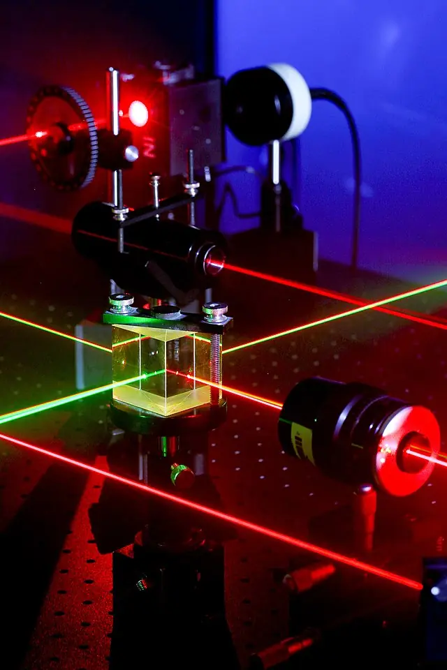
SLO is based on the principle of confocal microscopy, which uses a laser beam to scan a small area of the eye and create high-resolution images of the structures within that area. The laser beam is directed onto the retina and is reflected back by structures in the eye, such as the retinal pigment epithelium and the choroid. This reflected light is then captured by a detector, which creates a two-dimensional image of the scanned area. The laser used in SLO is a low-power beam that reduces the risk of phototoxicity and makes it well-suited for imaging patients with light-sensitive eyes.
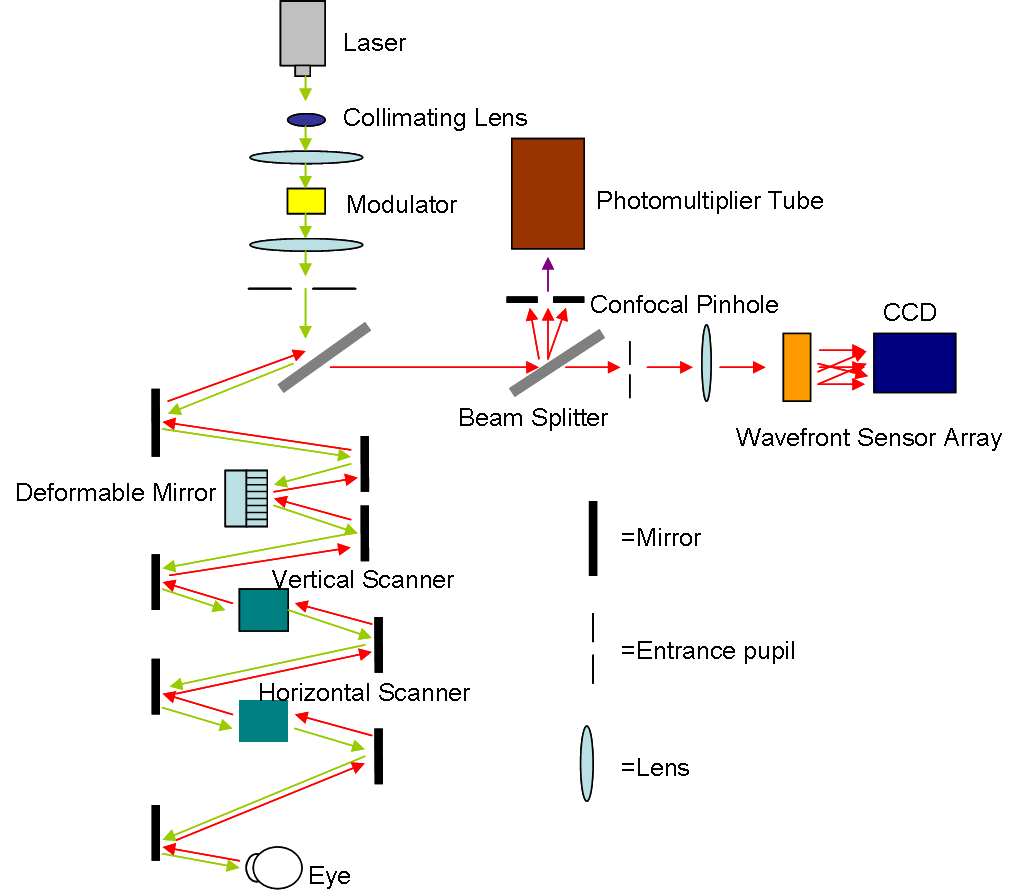
The SLO instrument consists of several key components, including a laser source, a scanning device, a detector, and a computerized system that processes the captured images. The laser source generates a low-power laser beam that is directed onto the retina. The scanning device, which can be a galvanometer or a resonant mirror, moves the laser beam across the retina in a raster pattern. The detector, which can be a charge-coupled device (CCD) or a photomultiplier tube (PMT), captures the reflected light and converts it into an electrical signal. The computer system processes the electrical signal and creates a high-resolution image of the scanned area.
SLO has several advantages over traditional ophthalmoscopy and fundus photography. One of the main advantages is its high resolution, which allows for the visualization of fine details in the retina and other structures. This high resolution is achieved by the confocal nature of the technique, which selectively captures only the light that is reflected from a specific depth within the eye. Additionally, SLO has a relatively low light level, which reduces the risk of phototoxicity and makes it well-suited for imaging patients with light-sensitive eyes.
SLO is used in the clinical setting for a wide range of applications. In the diagnosis and management of age-related macular degeneration, SLO can provide detailed images of the macula, including the presence and extent of drusen, which are the hallmark of the disease. In the management of diabetic retinopathy, SLO can be used to detect and monitor the progression of microvascular changes, such as leakage from blood vessels, which are indicative of the disease. In the management of glaucoma, SLO can be used to detect and monitor the progression of changes in the retinal nerve fiber layer, which is an indicator of nerve damage caused by the disease.
SLO is also a valuable tool for research in the field of ophthalmology. It provides high-resolution images of retinal structures, which can be used to better understand the underlying mechanisms of various eye conditions. This information can then be used to develop new treatments and therapies for these conditions. Furthermore, SLO can be used to evaluate the efficacy of existing treatments, allowing for ongoing monitoring of patients and refinement of treatments over time.
In conclusion, SLO is a highly advanced and non-invasive imaging technique that provides high-resolution images of the retina and other eye structures. Its ability to detect and monitor retinal changes and its low light level make it an invaluable tool in the diagnosis and management of eye conditions such as age-related macular degeneration, diabetic retinopathy, and glaucoma. SLO is a valuable addition to the ophthalmologist’s diagnostic arsenal and an essential tool in the field of ophthalmology.