How Multiline Illumination Confocal Raman Microscopy is Transforming Biomedical Research
Raman spectroscopy is a powerful and non-invasive tool that allows researchers to obtain important insights into the chemical makeup of a sample. This tool can be used to perform complex chemical analysis of cells and tissues, and it is becoming increasingly important in biomedical applications. In order to improve the usefulness of Raman spectroscopy in the clinic, researchers have developed a high-throughput Raman microscope that can acquire information hundreds of times faster than conventional Raman microscopes. This development could bring Raman spectroscopy closer to being a practical tool for medical diagnoses and drug screening.
The researchers from Osaka University have described their new multiline illumination confocal Raman microscopy approach in the Optica Publishing Group journal Biomedical Optics Express. This approach works by detecting separate regions of the sample in parallel, enabling fast Raman hyperspectral imaging. The team showed that the technique can acquire hyperspectral images of biological tissue with a field of view of 1380 x 800 pixels in just 11 minutes, a process that would take days to acquire with a traditional Raman microscope.
The new multiline illumination approach builds upon a technique previously developed by the research team known as line-illumination Raman microscopy. This approach was faster than conventional confocal Raman microscopy and enabled dynamic imaging of living cells, but it was still too slow for the large-area imaging often required for medical diagnosis and tissue analysis. The multiline illumination approach solves this issue by acquiring large-area images about 20 times faster than line-illumination Raman microscopy.
The team’s new multiline-illumination Raman microscope irradiates about 20,000 points in a sample simultaneously with multiple line-shaped laser beams. The Raman scattering spectra generated from these positions are then recorded in a single exposure that contains the spatial information for the Raman spectra in the sample. This information is then used to reconstruct a two-dimensional hyperspectral Raman image. The researchers use a cylindrical lens array, a custom-made spectrophotometer, optical filters, and a high-sensitivity, low-noise CCD camera with a large number of pixels to accomplish this.
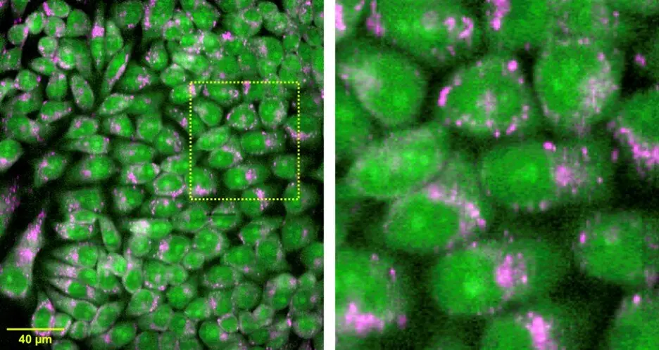
The researchers tested the new technique by acquiring measurements from live cells and tissues, including mouse brain, kidney, and liver tissue. They showed that the technique could acquire 1,108,800 spectra in just 11.4 minutes. The researchers believe that this technique could also be applied for small-molecule imaging and super-multiplex imaging using Raman tags and probes, as it benefits from fast imaging and does not require a large number of pixels in a spectrum.
In order for this technique to be applied for medical diagnoses, the researchers say it is important to build a database of Raman images. This can be accomplished efficiently with the new Raman microscope due to its speed and large imaging area. The researchers are also working to increase the system’s speed by a factor of about 10 and reduce the cost of the camera, laser, and spectrophotometer to make commercialization more practical.
In conclusion, the development of a high-throughput Raman microscope could bring Raman spectroscopy closer to being a practical tool for medical diagnoses and drug screening. The researchers’ multiline illumination confocal Raman microscopy approach provides fast and non-invasive imaging and analysis of biological tissue. The researchers believe that this technology could overcome the limitations of current methods and enable new applications in biomedical research.